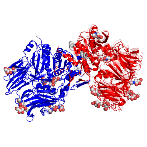
ID.47 |
DIPEPTIDYL PEPTIDASE IV |
|
|
|
Function |
EC*1 |
CSA distance*2 |
3.4.14.5 |
20.7 |
|
*1 Enzyme commission number. |
Ligand |
PDB*1 |
Full name |
11xNAG,5xNDG |
11x(N-ACETYL-D-GLUCOSAMINE),5x(2-(ACETYLAMINO)-2-DEOXY-A-D-GLUCOPYRANOSE) |
|
*1 Ligand name designated by the PDB identifiers. |
Segments |
Component No. |
Fixed*1 |
Moving*1 |
Motion type |
Ligand binding |
Coupled motion type |
1 |
D1(41A-236A,252A-546A,548A-735A,743A-761A,239B-255B,702B-702B,726B-731B,734B-741B,754B-756B) |
D2(237A-241A,244A-244A,248A-251A,736A-742A,762A-764A,39B-238B,256B-546B,548B-650B,658B-672B,674B-678B,690B-691B,699B-701B,703B-711B,716B-718B,720B-725B,732B-733B,742B-753B,757B-762B) |
Domain |
Independent |
|
*1 The location of the fixed and moving segments indicated by the residue number assigned in the ligand-bound form. The background color of characters indicates the corresponding segment in the structure. The colored segments not described in the Table are: 1) a part of component in which the motion is small (< 1.0 A), or, 2) a part of a protomer of homodimers, for which a corresponding part of the other protomer is shown in the Table. |
Displacement and disorder |
Component No. |
RMSD*1 |
Displacement*2 |
Disorder-order transition*3 |
Disorder residue*4 |
Helix-Coil*5 |
1 |
1 |
|
*1 The root-mean-square displacement of a component of motion calculated for the domain motions. |
Crystal environment |
Component No. |
Open state*1 |
Crystal packing*2 |
1 |
Bound |
Coupled |
|
*1 Distinction between the open state and the closed state required for examining the influence of the crystal environment. |

