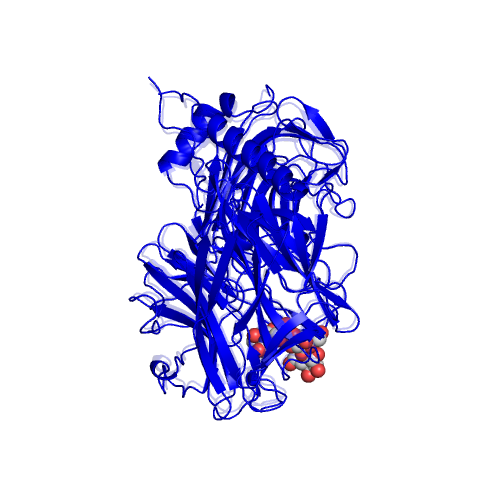
O.5 |
TRANS-SIALIDASE |
|
|
|
Function |
EC*1 |
CSA distance*2 |
3.2.1.18 |
|
*1 Enzyme commission number. |
Ligand |
PDB*1 |
Full name |
DAN,LAT |
2-DEOXY-2,3-DEHYDRO-N-ACETYL-NEURAMINIC ACID,BETA-LACTOSE |
|
*1 Ligand name designated by the PDB identifiers. |
Segments |
Component No. |
Fixed*1 |
Moving*1 |
Motion type |
Ligand binding |
Coupled motion type |
1 |
D1(3B-6B,10B-11B,234B-243B,246B-251B,253B-258B,263B-272B,274B-285B,288B-289B,303B-305B,309B-311B,316B-318B,327B-328B,333B-359B,362B-362B,365B-386B,398B-399B,409B-414B,419B-421B,434B-439B,467B-467B,474B-475B,482B-487B,500B-504B,513B-514B,549B-570B,580B-585B,606B-607B,609B-621B) |
D2(7B-9B,12B-14B,20B-20B,29B-39B,51B-61B,91B-91B,93B-98B,108B-112B,118B-122B,130B-135B,154B-174B,181B-209B,219B-233B,244B-245B,252B-252B,259B-262B,286B-287B,360B-361B,363B-364B) |
Other/ domain-like |
Coupled |
|
*1 The location of the fixed and moving segments indicated by the residue number assigned in the ligand-bound form. The background color of characters indicates the corresponding segment in the structure. The colored segments not described in the Table are: 1) a part of component in which the motion is small (< 1.0 A), or, 2) a part of a protomer of homodimers, for which a corresponding part of the other protomer is shown in the Table. |
Displacement and disorder |
Component No. |
RMSD*1 |
Displacement*2 |
Disorder-order transition*3 |
Disorder residue*4 |
Helix-Coil*5 |
1 |
1 |
|
*1 The root-mean-square displacement of a component of motion calculated for the domain motions. |

