O.8 |
HYPOXIA-INDUCIBLE FACTOR 1 ALPHA INHIBITOR |
|
|
|
Function |
EC*1 |
CSA distance*2 |
1.14.11.16 |
|
*1 Enzyme commission number. |
Ligand |
PDB*1 |
Full name |
2xPEPT(QLTSYDCEVNAPI),2xZN,2xAKG |
2x(Peptide(QLTSYDCEVNAPI)),2x(ZINC ION),2x(2-OXOGLUTARIC ACID) |
|
*1 Ligand name designated by the PDB identifiers. |
Segments |
Component No. |
Fixed*1 |
Moving*1 |
Motion type |
Ligand binding |
Coupled motion type |
1 |
D1(347A-349A,12A-13A,37A-40A,52A-54A,57A-60A,97A-120A,195A-200A,215A-219A,224A-260A,274A-279A,340A-342A) |
D2(33A-36A,72A-96A,121A-147A,155A-155A,157A-157A,164A-164A,191A-194A,212A-214A,280A-282A) |
Other/ domain-like |
Coupled |
|
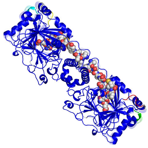
*1 The location of the fixed and moving segments indicated by the residue number assigned in the ligand-bound form. The background color of characters indicates the corresponding segment in the structure. The colored segments not described in the Table are: 1) a part of component in which the motion is small (< 1.0 A), or, 2) a part of a protomer of homodimers, for which a corresponding part of the other protomer is shown in the Table. |
Displacement and disorder |
Component No. |
RMSD*1 |
Displacement*2 |
Disorder-order transition*3 |
Disorder residue*4 |
Helix-Coil*5 |
1 |
1 |
|
*1 The root-mean-square displacement of a component of motion calculated for the domain motions. |

