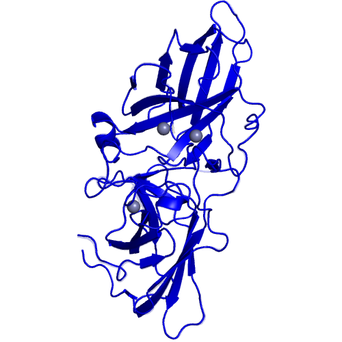
B.65 |
34 KDA MEMBRANE ANTIGEN |
|
|
|
Ligand |
PDB*1 |
Full name |
3xZN |
3x(ZINC ION) |
|
*1 Ligand name designated by the PDB identifiers. |
Segments |
Component No. |
Fixed*1 |
Moving*1 |
Motion type |
Ligand binding |
Coupled motion type |
1 |
Buried ligand |
|
*1 The location of the fixed and moving segments indicated by the residue number assigned in the ligand-bound form. The background color of characters indicates the corresponding segment in the structure. The colored segments not described in the Table are: 1) a part of component in which the motion is small (< 1.0 A), or, 2) a part of a protomer of homodimers, for which a corresponding part of the other protomer is shown in the Table. |
H-bond |
Component No. |
Ligand*1 |
Water*1 |
Protein*1 |
1 |
14 |
5 |
3 |
|
*1 The number of hydrogen-bonds observed in the proteins classified in "Buried Ligand". |

